Category Archives: News and Press

AOC PATTON BLOG Articles

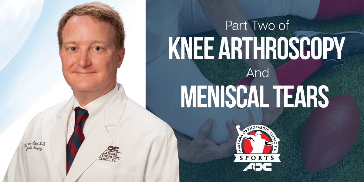
Knee Arthroscopy and Meniscal Tears – Part Two

The treatment of meniscal tears has advanced over the years. Forty years ago it would not have been unheard of to excise in an open fashion the entire meniscus. This led to the development of arthritis earlier than was expected. With the development of knee arthroscopy and the small instruments to work inside the knee, debrided (shaving) just the tear became more common. This allowed meniscal tissue to be preserved.

As studies have shown the benefits of preserving meniscal tissue, repairing the meniscus, if possible, has become a goal. Several techniques including “outside-in”, “inside-out”, and “all inside” were developed. The term “all-inside” means the stiches are tied or the device deploys inside the knee without the need for additional incisions. This is also known as “all arthroscopic.” Regardless of the technique, the goal is to repair the meniscus in a stable manner so that early range of motion and healing are possible.

The indications for repair continue to expand with tears greater than one centimeter in the area of good blood supply and in patients less than 40 being common. Additionally, repairs at the time of ACL reconstruction, vertical tears, and acute tears also being good candidates for repair. As the technology improves the candidates for repair are expanding.

When meniscal tissue is severely lost in young patients with good alignment, intact ligaments, and no significant arthritis, meniscal transplant has been developed as an option. Taking a meniscus from a young donor and either suturing it in the knee with or without a piece of bone attached can restore needed meniscus tissue. Long term studies of meniscal transplant are on-going. Hopefully as studies are produced and technology develops, preservation of meniscal tissue will continue to advance in the hope of preserving the articular cartilage and normal biomechanics of the knee.
AOC PATTON BLOG Articles

Knee Arthroscopy and Meniscal Tears – Part One


Knee arthroscopy (the placing of a small camera into the knee and performing surgery through small holes) is one of the most common orthopaedic procedures performed in the United States. Over 900,000 of these are performed each year, and over half are done to operate on a torn meniscus.1 The meniscus is a C-shaped cartilage structure on the inside (medial) and outside (lateral) of the knee. Acute traumatic tears of the meniscus are often caused by sporting activities or an activity where there is a sudden twisting of the knee. Hyperflexion of the knee can cause meniscal tears also. Degenerative type tears can be caused by age or untreated instability of the knee.

The meniscus serves an important function in the knee by acting as a cushion to the underlying joint cartilage called the articular cartilage. Loss of meniscus tissue through tearing or surgical removal can lead to increases in stress placed upon the articular cartilage. This can lead to arthritis. Additionally, the meniscus acts as a joint stabilizer. Loss of the meniscus can place increased stress on the major ligaments of the knee such as the anterior cruciate ligament (ACL). Therefore, preserving as much meniscus as possible is a goal for orthopaedic surgeons.

The diagnosis of a meniscal tear requires taking a history, performing a physical exam of the entire knee and surrounding structures, and performing additional diagnostic tests. As stated above, a history of a twisting injury or hyperflexion injury of the knee can lead to a meniscal tear. Common physical exam findings include swelling of the knee, joint line tenderness on the side of the tear, and pain with certain maneuvers that the physician performs such as a McMurray’s test. (The McMurray’s test is performed by taking the knee from a flexed position to an extended position while the tibia is kept internally rotated (for the lateral meniscus) or externally rotated (for medial meniscus). McMurray described a palpable click as being “positive”.2

Diagnostic tests for evaluating a torn meniscus include X-rays of the knee, MRI and knee arthroscopy. The plain X-rays do not detect the torn meniscus, but they rule out other causes of knee pain such as loose bodies and arthritis. The most common non-surgical diagnostic test for meniscal tears is the MRI. It has a high sensitivity and does not involve radiation. MRI can detect both meniscal and ligamentous tears. The “gold standard” test for diagnosing meniscal tears is actually seeing it with knee arthroscopy. This common outpatient procedure can be performed under general or spinal anesthesia.


1Kim et al: Increase in outpatient knee arthroscopy in the United States: a comparison of National Surveys of Ambulatory Surgery, 1996 and 2006. J Bone Joint Surg Am. 2011 Jun 1;93(11):994-1000.
2 McMurray TP: The semilunar cartilages. Br J Surg 1942;29(116):407–414

AOC-Barber-BLOG-Articles-FB-RoboticHipReplacement

The Advantages Of Robotic Hip Replacement

The Advantages Of Robotic Hip Replacement

Decades of experience and published studies have shown that the key to a long-lasting, well-functioning hip replacement is proper placement of the implanted device. Ensuring appropriate position and orientation of implants used to rely only on the skill of the surgeon. Now, many of us are employing technologies such as robotics to augment our skills and achieve better outcomes for patients. With the advent of robotic assisted surgery, we are able to create a 3D model of each patient’s anatomy and determine BEFORE surgery the best size of implants and how they should be positioned. DURING surgery, a robotic arm ensures that the surgery is carried out as close to the pre-operative plan as possible. While the goals of surgery have not changed, technology has improved the precision and accuracy with which we are able to perform it. This increased precision is all aimed at getting the best and most durable result that can possibly be achieved for each patient.

For more information, please visit: barbertotaljoint.com


AOC-Barber-BLOG-Articles-3D-Knee

What Is A Custom 3D Knee Replacement?

Dr. Barber answers the question - What Is A Custom Knee Replacement 3D Knee?

In recent years there has been great interest in custom implants for total knee replacements and I am asked about them frequently. These are sometimes referred to as “the 3D knee” because they are produced using 3D printing technology. With this system, a CT scan is used to produce custom instrumentation and a custom prosthesis which are used in surgery. Each implant is truly one of a kind. These devices are intended to address some common problems in knee replacement such as size mismatch or improper position. Because the implants are created specifically for each patient’s anatomy there is increased potential for the replaced knee to function more like the patient’s natural knee. Over the last few years, our experience with these implants has been truly remarkable. To see if you are a candidate for this procedure, contact us at AOC Joint Replacement and schedule an appointment today.
For more information, please visit: mobilekneesurgeon.com


AOC-Barber-BLOG-Articles-You-knee-que

U Are You-KNEE-que

Dr. Barber talk about how each patient’s knee problems are unique and specific

Each patient’s knee problems are unique and specific. When choosing your care it is important to find a fellowship trained surgeon who specializes in your condition. I have produced great results with partial knee replacements, customized knee implants,  and robotic surgeries; but there is no “one size fits all” solution for knee problems. Every patient deserves a thorough evaluation and a solution which matches their lifestyle and expectations. I always work with a patient to determine what their You-KNEEque needs are and how we can meet them.

For more information, please visit: mobilekneesurgeon.com


AOC-Barber-BLOG-Articles-FB-Knee-Pain

Knee Pain? Partial Knee Replacement Might Be An Option

Dr. Barber, Knee Pain? Partial Knee Replacement  in Mobile AL

Many people suffering from knee pain avoid seeking help because they fear being told that they will require total knee replacement. Often there is a better option for patients living with arthritis pain, but wishing to remain active. Partial knee replacement is a less invasive surgery with a much quicker recovery that can aid in restoring normal function. These procedures are often performed on an outpatient basis. Because all of the knee ligaments are retained, the motion of a partial knee replacement is typically greater and the mechanics are remarkably similar to that of a normal knee. Determining if a partial knee replacement is the right choice requires a careful evaluation by a surgeon who is well-versed in all of the options available and can guide you to the best treatment for YOUR knee.
For more information, please visit: mobilekneesurgeon.com


Dr. Donahoe explains Lumbar Fusion when spinal fusion is used to stabilize the spine when arthritis has led to instability of the spinal segment.

Lumbar Fusion

AOC-Donahoe-BLOG-Articles-WEB-Lumbar-Fusion

Have you been diagnosed with a slipped disc in your back and your doctor recommended surgery? Spinal fusion is a procedure to stabilize the spine when arthritis has led to instability of the spinal segment (2 bones around the disc). This procedure is usually done along with a laminectomy to relieve the pressure on the spinal nerves. Patients with mechanical low back pain and pain in the legs when standing or walking, may benefit from this procedure. Screws and rods are placed around the spine for stability along with bone graft for the fusion.
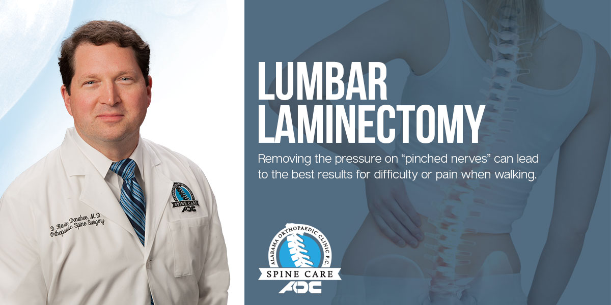


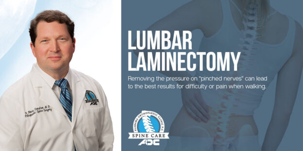
Dr. Donahoe explains lumbar laminectomy and how the best results come from removing the pressure on “pinched nerves” in the neck or back.

Lumbar Laminectomy

AOC-Donahoe-BLOG-Articles-FB-Lumbar-Laminectomy

Have you been diagnosed with spinal stenosis? Do you have difficulty walking any long distances without experiencing leg pain? Patients who have not improved after conservative treatment may benefit from surgery. During surgery, the bone spurs and arthritis are removed that place pressure on nerves that affect the legs and buttock region. Dr. Donahoe specializes in lumbar laminectomy surgery and would be happy to evaluate your MRI. Like most spine surgeons, the best results come from removing the pressure on “pinched nerves” in the neck or back.


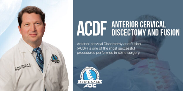
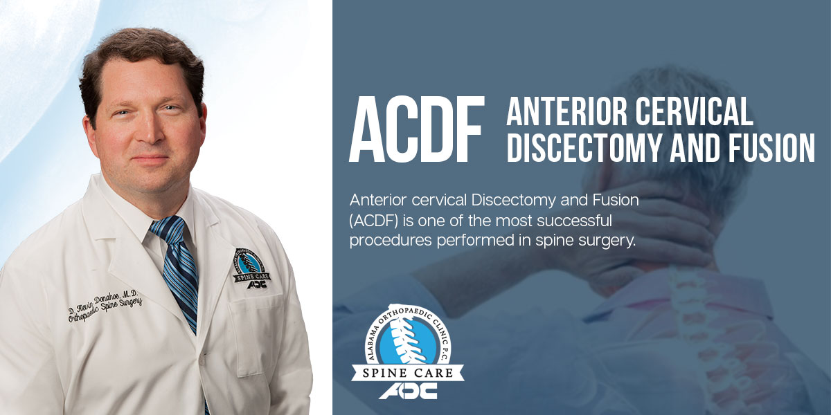
Dr. Donahoe explains why Anterior Cervical Discectomy and Fusion (ACDF) is one of the most successful procedures performed in spine surgery.

Anterior Cervical Discectomy and Fusion (ACDF)

Dr. Donahoe explains Anterior cervical Discectomy and Fusion (ACDF) is one of the most successful procedures performed in spine surgery.

Have you been diagnosed with a pinched nerve in your neck? This condition can cause pain that radiates to the shoulder or arm. Patients who have not improved with conservative treatment including medication and physical therapy may be candidates for surgery. Patients typically spend one night in the hospital, and almost immediately after surgery are relieved from the arm pain, numbness, and tingling.  Anterior Cervical Discectomy and Fusion (ACDF) is one of the most successful procedures performed in spine surgery.